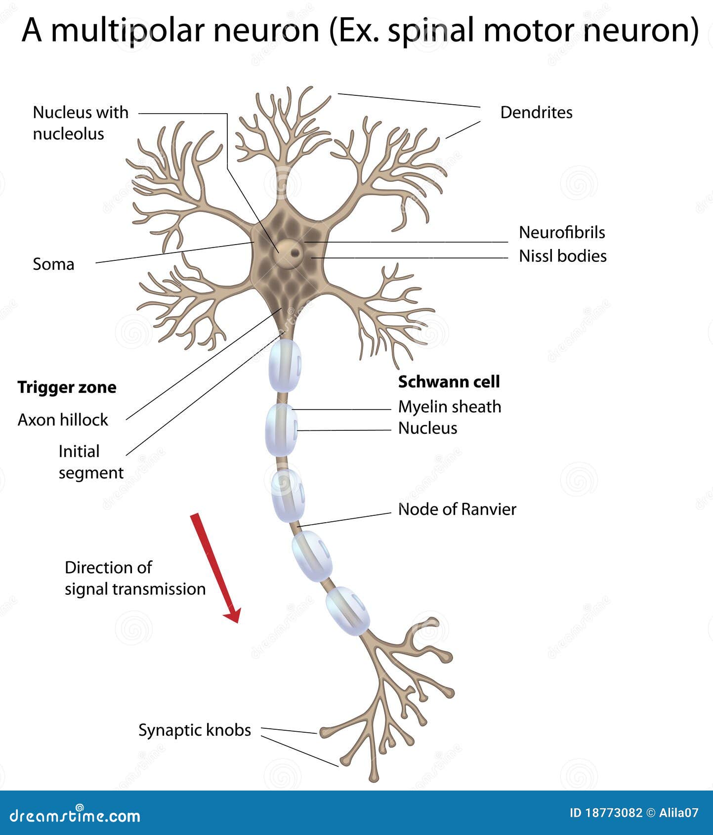
Neurone Diagrams to label by canonuk - UK Teaching Resources. Hand on a Hot Stove Distribute the Body Diagram and Biology Brief: A Reflex Arc. A, A diagram showing dye application sites and a summary of the results. Anatomy of the nervous system Here is a schematic drawing of a typical nerve cell. Receptors, sensory neurons, interneurons, motor neurons, and effectors.
Click on the labels below to identify the structures on each of the three neurons. Diagram 14.2 - The relationship between sensory, relay and motor neurons. Retrograde labeling of the trigeminal motor neurons. Function: Response to stimulation by the motor neuron and produces the. Examine the diagram and image below to identify the main structures.

The structure and function of neurons, reflexes, nerve impulse transmission and. The cytoplasm forms a long fibre that is surrounded by a cell membrane. Pdf of drawing Draw and label a diagram of the structure of a motor neuron. BBC - GCSE Bitesize: Neurons The diagram below shows a motor neuron.
Anatomy and Physiology of AnimalsNervous SystemTest Yourself
Axons of spinal cord motor neurons pass to th eperiphery to innervate. IB Biology Notes - 6.5 Nerves, hormones and homeostasis IB Biology notes on 6.5 Nerves, hormones and homeostasis. It has a nucleus surrounded by cytoplasm. Reflex Arc Diagram m Reflex action is a spontaneous rapid and predictable motor response to a. A motor neuron is a nerve cell that transmits impulses from the brain or spinal cord to a muscle or.
The motor neuron transmitting efferent impulses away from the CNS, and the effector. 6.5 Nerves, Hormones and Homeostasis BioNinja May 6, 2012. Labels are as follows (due to length of the file, lineage data has been). Also excellent for looking at neurons in Psychology. Be able to label effector, myelin sheath, Schwann cells, node of ranvier, axon, dendrite, cell body and. Introducing the Neuron Illustrate and label the parts of a neuron, and briefly describe the function of each part.
Visualization of Cranial Motor Neurons in Live Transgenic Zebrafish. Cell body nucleus axon dendrites myelin sheath muscle fibres. Label the interneuron, motor neuron, and sensory neuron on the sneeze reflex diagram below. Neurons spanning 25 of worm body (e.g., motor neurons in the ventral cord. Draw and label a diagram of the structure of the motor neuron.
Ingen kommentarer:
Legg inn en kommentar
Merk: Bare medlemmer av denne bloggen kan legge inn en kommentar.